I’ve talked about protein structure alignment before in the context of a rather novel, mathematical approach. This time I wanted to revisit the topic in a general sense, using a more established algorithm as a case study. MAMMOTH stands for Matching Molecular Models Obtained from Theory and was first published in 2002. Since then it has been cited nearly 400 times and the underlying algorithm has been extended to a multiple alignment program: MAMMOTH-mult.
Establishing biologically relevant and consistent alignments between protein structures is one of the major unsolved problems in computational bioinformatics. However, it’s an important part of many challenges that we face: such as establishing homology between distantly related proteins, functional inference for unannotated proteins, and evaluating the accuracy of models of predicted structure for competitions such as CASP.
Problem Outline
In essence the problem of protein structure alignment can be outlined by considering two ordered sets of coordinates, A = {a1,a2,…,an} and B = {b1,b2,…,bm}, representing points in 3D space. In most cases these points will be the location of the Cα atoms along each structure’s backbone. The sets A and B might be completely different lengths and, if an alignment exists, are almost certainly orientated differently compared to each other.

Establishing an alignment between these sets is equivalent to two steps:
- Establish a match M = {(ai,bj) | ai ∈ A, bj ∈ B}
- Rotate and translate A onto B so that equivalent atoms are as close as possible.
Of course, it is not immediately clear how to best fulfill these requirements. In particular, we don’t really know what features to prioritise for a biologically relevant match. Should we try to match secondary structure elements and what penalty should we attach to mismatched elements? How about maintaining the correct hydrogen bonding patterns between matched residues? And how much weight should we put on the matched atoms being consecutive in each set (i.e. how should we penalise gaps)?
The second step is equally ambiguous. Especially as there is no consensus on what the correct interpretation of close is. Minimising the RMSD between equivalent atoms is a popular choice of distance measure. However, as the MAMMOTH paper points out, RMSD is often dominated by the mismatched portions of remotely related structures and is thus largely inappropriate in these cases. Furthermore, even if we have a well-defined distance metric, should the superposition prioritise minimising the distances between nearly identical parts of the different structures, at the expense of less similar substructures? Or should the emphasis be on maintaining as lengthy a match as possible at the possible cost of a lower closeness of fit? How about the relative importance of a close fit for atoms in the core of the structure vs. those on the surface?
The majority of these questions remain unanswered and as a result it is often hard to validate alignments as we simply do not know what the right answer is. In fact, in many cases, manual analysis is preferred over any of the available computational techniques.
In this post I’ll go through how the MAMMOTH algorithm approaches each of these steps. For many of the above questions MAMMOTH does not postulate a solution, primarily because, as its name suggests, it was designed to assess prediction models which are often at low resolutions and lacking secondary structure or hydrogen bonding information. I think it’s important to keep these questions in mind, but there’s certainly no necessity to design a programme which deals with them all.
Step 1: Pairing up residues (similarity matrix)
In order to establish a match between equivalent atoms in A and B, MAMMOTH, like several other structural alignment algorithms, uses a well-established alignment technique: a similarity matrix (often inferred from and referenced as a distance matrix). A similarity matrix for alignment is an n x m array where each entry S(ai,bj) represents the pairwise similarity score between residues ai and bj. An alignment between the residues of A and B is any non-decreasing path (that is, a pair (ai,bj) in the path must appear later in the ordering of coordinates of both A and B than the preceding pair of residues in the path) from the top left corner of the array (a1,b1) to the bottom right corner (an,bm). For example the following path can be interpreted as an alignment between A = {a1, …, a11} and B = {b1, …, b8}

Any alignment can be scored by summing up the similarity scores along this path, while penalising any gaps in an appropriate way (normally, these algorithms use trial and error to decide on sensible penalties). For example, the above alignment would have the score S = S(a1,b1) + S(a2,b2) + S(a3,b3) + S(a7,b4) + S(a8,b5) + S(a9,b6) + S(a10,b7) + S(a11,b8) + α + 2β, where α and β are gap opening and gap extension penalties respectively. The optimal alignment is simply the alignment which maximises this score.
For sequence alignments similarity scores can be assigned to residues from substitution tables like BLOSUM. However, it is not immediately clear of an appropriate equivalent for structures. MAMMOTH, like several other algorithms, defines the similarity between different residues by examining their local structural landscape. Specifically, this means comparing fragments of each backbone, centred on the residue of interest. MAMMOTH uses the URMS distance between heptapeptide fragments. This distance is illustrated below using 2D chains and tripeptide fragments.

Comparing residues a2 and b3 involves looking at the directions between each successive residue of the fragment. Each vector is mapped to the unit sphere, beginning at the origin and ending at the surface of the sphere (in this case 2 vectors are considered, and in MAMMOTH’s case 6 different 3D vectors are mapped). The optimal rotation is found, superposing equivalent vectors as best as possible, and then the RMSD of the endpoints on the surface of the sphere is calculated as URMS(ai,bj).
Aside: The optimal superposition of a set of vectors is actually a non-trivial problem. It is essentially equivalent to step 2 in our alignment protocol outlined above, but is significantly easier for the 6 vectors characterising a fragment in MAMMOTH’s algorithm.
Finally, S(ai,bj) is calculated by converting the distance into a similarity measure:

where URMSR is the expected URMS of a random set of vectors and:

The optimal alignment through this MAMMOTH matrix is the path which maximises the sum of similarities between matched residues (each residue being at the centre of the heptapeptide fragment) using gap opening and extension penalties of 7.00 and 0.45 respectively.
Step 2: Global superposition (MaxSub)
The above alignment results in a match M’ optimising the local structural similarity of residues in each structure, however, their is no guarantee that this will result in a set of coordinates close in global space. In order to finalise the match set M ⊆ M’ as well as calculating the optimal superposition of the paired residues of A onto their equivalent points in B, MAMMOTH use the MaxSub algorithm. This is a very efficient algorithm (worth a read if you’re that way inclined) for calculating the maximal subset from a set of paired up atoms which are close in global space. MAMMOTH decide that close means < 4A away after superposition. They do not try to optimise a closer superposition than that but attempt to find the largest possible set of matched residues.
The MaxSub algorithm relies on the assumption (made for computational tractability) that the final subset M ⊆ M’ will have, somewhere, a set of at least four residues consecutive in M’. The algorithm then starts with every possible seed of four consecutive residues (just to illustrate the power of the assumption in reducing computational time: for a 150 residue protein there are just 147 such seeds, but over 2 million sets of four non-consecutive residues!! And it’s a pretty reasonable assumption to make as well). The MaxSub algorithm then calculates the superposition for those four matched pairs, extending the set of residues that are <4A away from their partners, recalculating the superposition using these new pairs as well, then removing any pairs which are no longer within the threshold of each other. It repeats these steps, gradually extending the set M, until the algorithm converges.
Scoring the alignment
Using the two approaches outlined above, MAMMOTH generates an alignment between the two input structures. In order to summarise the significance of this alignment, the algorithm generates the PSI score: the percentage structural identity (which is simply the size of the maximum subset divided by the length of the shortest protein). As a global measure of the strength of similarity the PSI score is poorly constructed and scales with protein length. In order to adjust for this bias, MAMMOTH fits a Gumbel distribution to PSI scores obtained from random structure comparisons between unrelated proteins at bins of different lengths. This results in a z-score measuring, instead of the PSI of an alignment, the likelihood of obtaining a PSI score as good as that by chance between any two proteins of the same lengths.

 Identifying the Correct Model
 . In this article they proposed an exponential family of probability distributions to model
  , where
  is a possible realisation of the random matrix
  ) (see nodes 3-5 of the Figure below).


 are parameters, and
  (identifying constrains).
  can be interpreted as the mean tendency of reciprocation,
  can be interpreted as the density (size) of the network,
  can be interpreted as as the productivity (out-degree) of a node,
  can be interpreted as the attractiveness (in-degree) of a node.
 and
and  are: the number of reciprocated edges in the observed graph, the number of edges, the out-degree of node i and the in-degree of node j; respectively.
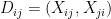 , the model assumes that all
  with
with  are independent.
 and describe the joint distribution of
  , assuming all


 .
.
 for
for  , and
, and 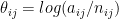 for
for  .
. and
and  can be interpreted as the reciprocity and differential attractiveness, respectively. With a bit of algebra we get:
![exp(\rho_{ij})=[ P(X_{ij}=1|X_{ji}=1)/P(X_{ij}=1|X_{ji}=0) ]/[ P(X_{ij}=1|X_{ji}=0) / P(X_{ij}=0|X_{ji}=0) ],](https://s0.wp.com/latex.php?latex=exp%28%5Crho_%7Bij%7D%29%3D%5B+P%28X_%7Bij%7D%3D1%7CX_%7Bji%7D%3D1%29%2FP%28X_%7Bij%7D%3D1%7CX_%7Bji%7D%3D0%29+%5D%2F%5B+P%28X_%7Bij%7D%3D1%7CX_%7Bji%7D%3D0%29+%2F+P%28X_%7Bij%7D%3D0%7CX_%7Bji%7D%3D0%29+%5D%2C+&bg=ffffff&fg=000&s=0&c=20201002)

 , and
, and  where
where 


