
SAbDab has been around since 2013. Its now over 22,000 antibody structures have been searched and downloaded and searched again by a great many people with a great many browser tabs open, clicking, and scrolling, and clicking away. Starting this week, they no longer have to.
SAbDab2 now has a public API. You could read all about it in the documentation—or you could be sensible and just point your agent at this. Ready for a demo? I know I am.
What’s the most-solved antibody?
Wonder no more.
Hey, Claude, which antibody has the most structures in SAbDab2? Show me every one of them, and what it's bound to.
Two flibbertigibbets later, we have this:

24 selected GPCR complexes (of 552), coloured by family, superposed on the single scFv16 (blue) that holds their G protein (grey) in place.
Well, now you know. It’s not trastuzumab, for once. scFv16 is present in 552 PDB entries—just about all of them cryo-EM models. Its job is to hold GPCRs steady during imaging. The actual receptor changes from paper to paper.
What makes this query possible is SAbDab2 IDs.
Every distinct variable domain gets one stable identifier, based on its IMGT sequence numbering.
scFv16, for example, is sabdab2_H01TTL01FP.
That’s heavy chain H01TT and light chain L01FP.
Yes, we do think our naming scheme is an improvement over ‘scFv16’, why do you ask?
Every structure of it is filed under that ID, even if it were solved as an Fv or Fab.
Find everything that binds TNF-α, and show me where.
Right, you do actual work. Valid. You do you.
Find every antibody in SAbDab2 against human TNF-α. How many distinct antibodies is that, and in what formats are they available? Figure out where each one binds based on its best-resolved structure.
“Format” here is what’s in the crystal, not necessarily the vial. SAbDab2 now automatically recognises and annotates a range of novel antibody formats and constructs, including scFvs, diabodies, and more.
OK, it pulled the antigen sequence from UniProt… Querying SAbDab2… Three, two, one, done.

All twelve distinct antibodies (SAbDab2 IDs) against TNF-α, each from its best-resolved TNF-bound structure, placed on the same TNF-α trimer (4G3Y). TNF-α residues within 4.5 Å (heavy atoms) of the antibody highlighted; the darker protomer is the one it touches most. Canine-TNF complex drawn on human TNF-α. Ozoralizumab is the only ‘construct’ (a trivalent, chain-sharing nanobody). Constant domains are trimmed throughout.
Nice. Found the one against the dog homologue, too (94% identical). Ooh, and VNARs, really?!, interesting…
Maybe not the most useful view, though. Could we get that as a …

TNF-α’s most-targeted surface: each residue coloured by how many of the 12 distinct antibodies touch it. Counts are per residue position, pooled over the three identical subunits, and painted on all three.
…heatmap, yes, thank you, Claude. Looks like most antibodies bind across the groove between subunits of the homotrimer.
Right then, that took us a single prompt and, what, minutes? I have half a blog post left to write; let’s try something slightly more complex.
Tell me which EGFR antibodies might work in mouse.
I work in mouse models. My target is EGFR (mouse: UniProt Q01279). Do we have any structures of antibodies against that?
Turns out: no. Can’t have everything.
Find every antibody structure in SAbDab2 against human EGFR, map each one's epitope, and tell me how conserved it is between human and mouse. Do we have any antibodies that might cross-react?
Puttering, pondering, percolating, … presto.
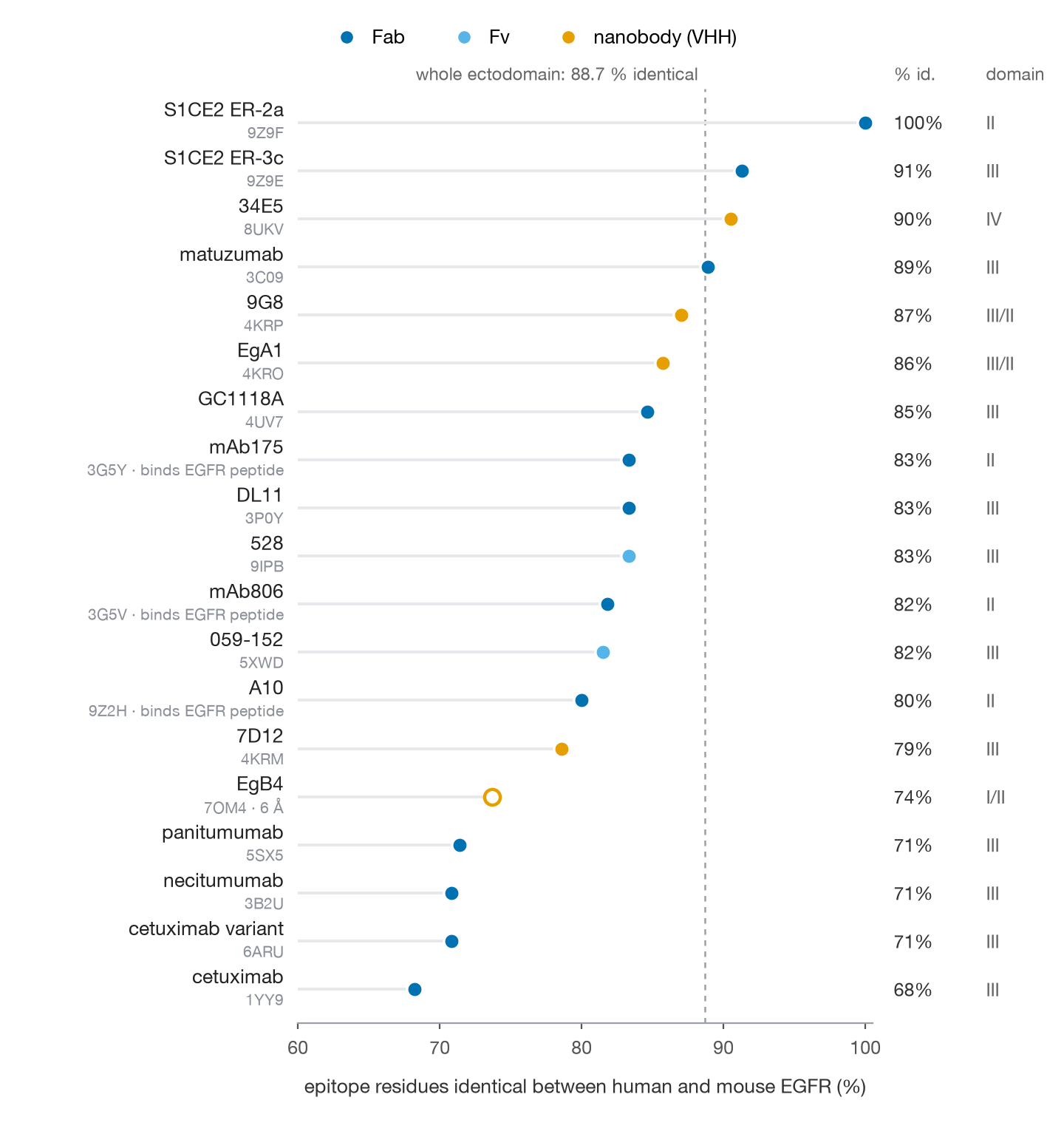
Epitope conservation for every SAbDab2 ID bound to wild-type EGFR. The hollow marker is a low-resolution structure (6 Å).
Well, that first one looks promising, as long as we’re OK with domain II.
What does S1CE2 ER-2a look like on the structure and what resolution is that?

S1CE2 ER-2a (sabdab2_H0642L04K0) on the human EGFR ectodomain, as captured in its complex structure (9Z9F, 3.1 Angstrom). Epitope residues blue: all 24 on the domain II dimerisation arm, and identical in mouse EGFR.
Looks reasonable, I guess.
Give me the CDRs.

CDR sequences of S1CE2 ER-2a, as annotated in SAbDab2. Numbering and CDR definitions follow IMGT conventions.
Find the most similar murine scaffold and swap them in...
… you get the idea. With a little bit of luck, we’ve got ourselves a mouse antibody, no de novo design required (pending the small matter of actually testing it).
Tell me what’s new this week.
Can’t keep up with the literature?
No one can.
SAbDab2 updates weekly and the number of antibody structures available is rising ever faster.
Ask your agent what’s new this week and you’ll get something like this:
32 new PDB entries, 35 distinct antibodies (29 new to SAbDab2). Firsts for the database include the orphan GPCR GPR55, fibroblast activation protein, interferon-α8, CD84 and a prefusion EBV gB.

Structure of the week: 9Y5C, the chimeric antibody 2C7 bound to a gonococcal lipooligosaccharide at 1.6 Å, a carbohydrate epitope on a bacterium that is outrunning our antibiotics.
Schedule that as a weekly job and you have a newsletter. Alternatively, describe what you’re interested in (an antigen, a format, an organism) and ask it to alert you whenever a new matching structure becomes available.
Get me clean data for ML.
Oh, did we mention? We’ve also cleaned up a bunch of high-quality antibody structures for you, so you can train models on them out of the box. We encourage you to use our pre-made splits. Find out all about them and get the data on Zenodo. Updated every six months.
What else?
None of this works without the curation underneath; a lot of painstaking quality control and manual annotation; and so, so much work on the backend. With SAbDab2, we have re-written our entire processing pipeline from scratch, improved and corrected annotations at every step, and are now able to process many more structures than ever before. We describe exactly how everything works—and give a full comparison to legacy SAbDab—in our paper, recently published in the NAR Database Issue.
If you’d rather look than read, you can browse SAbDab2 yourself, or simply install our live desktop wallpaper, and let corporate-chic antibody structures come to you. 😉

The wallpaper in dark mode. It pulls a random antibody–antigen complex from SAbDab2 every few minutes and renders it with Mol* live on your desktop, making sure to rotate and zoom aesthetically. The structure shown is pdb_00007k22: five Fvs in complex with polyomavirus capsid protein 1. CDRs in red.
Get started.
- Visit SAbDab2 on the web.
- Access the API:
- Get data for ML.
- Read our paper.
All data are CC BY 4.0 and there are currently no rate limits. We can’t wait to see what you do with it.


