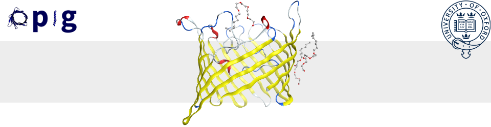
PyMOL is a handy free way of viewing three dimensional protein structures. It allows you to toggle between different representations of the protein – such as cartoon, surface, sticks, etc. – which all have their own pros and cons.
However one thing I felt that PyMOL lacked was an easy way to visually distinguish residues based on type. Whist you can easily differentiate atom types based on colour in the colour menu, and even choose which colour you wish carbons to show up as whilst keeping heteroatoms different colours, this assigned carbon colour would be constant throughout the entire protein.
Category Archives: Protein Structure
Cryo-EM reaches atomic resolution in protein structure determination
The end of the era of crystallography has been long predicted also on this blog, but has it come now with the recent report of an atomic resolution protein structure solved by cryo–electron microscopy (cryo-EM)?
Continue readingSpeaking about Sequence and Structure at a Summit
A couple of weeks ago I was lucky enough to be asked to speak at the 5th Computational Drug Discovery & Development for Biologics Summit. This was my first virtual conference – it was a shame I didn’t get to visit Boston, and presenting to my empty room was slightly bizarre, but it was great to hear what people have been working on, and there’s definitely something to be said for attending a conference in fluffy socks…

Smaller and Smaller Fragments
Fragment-based drug discovery (FBDD) is based on the idea that using small (< 300 Da), highly soluble compounds to screen against a target will give higher hit rates and sample chemical space more efficiently compared to screens using larger, drug-like compounds.
Constrained docking for bump and hole methodology
Selectivity is an important trait to consider when designing small molecule probes for chemical biology. If you wish to use a small molecule to study a particular protein, but that small molecule is fairly promiscuous in its binding habits, there are risks that any effects you observe may be due to it binding other proteins with similarly shaped binding pockets, instead of your protein of interest.
Continue readingIt’s been here all along: Analysis of the antibody DE loop
In my work, I mainly look at antigen-bound antibodies and this means a lot of analysing interfaces. Specifically, I spend a lot of my time examining the contributions of complementarity-determining regions (CDRs) to antigen binding, but what about antibodies where the framework (FW) region also contributes to binding? Such structures do exist, and these interactions are rarely trivial. As such, a recent preprint I came across where the authors examined the DE loops of antibodies was a great motivator to broaden my horizons!
Continue readingWhat can you do with the OPIG Antibody Suite? v2.0
Since my last blogpost on this topic back in 2018, OPIG has expanded its range of tools for antibody/BCR analysis. Here is an updated summary of the OPIG antibody databases and immunoinformatics tools.
Continue readingAntibody-protein binding and conformational changes
I came across a recent paper on the antibody-protein binding and conformational changes. As I work mainly on the binding site/Fv regions of antibodies, I am intrigued to see the role of the constant domains in the overall antibody function.
Continue readingReal Space Correlation Coefficient
Introduction
In crystalography we are often faced with the question of how well a part of our model fits the data. Now crystalography has well developed probability models for the reflection amplitudes given then entire fitted model, but these do not provide a metric for “how much of the ligand is inside the blob”. This is because the reflection based models are inherently global.
Continue readingProCare: cavity similarity searching and its applications to fragment-based drug design
ProCare [1] is a package developed at the University of Strasbourg which is able to align and score the similarity of protein cavities. The aim is to find ligand binding sites between different proteins that are similar enough to bind the same ligand. The method used in ProCare is designed to look particularly at fragment (~⅓ size of a druglike ligand) binding sites. The aim is to predict potential fragment hits by comparing the cavities of the targets.
Continue reading